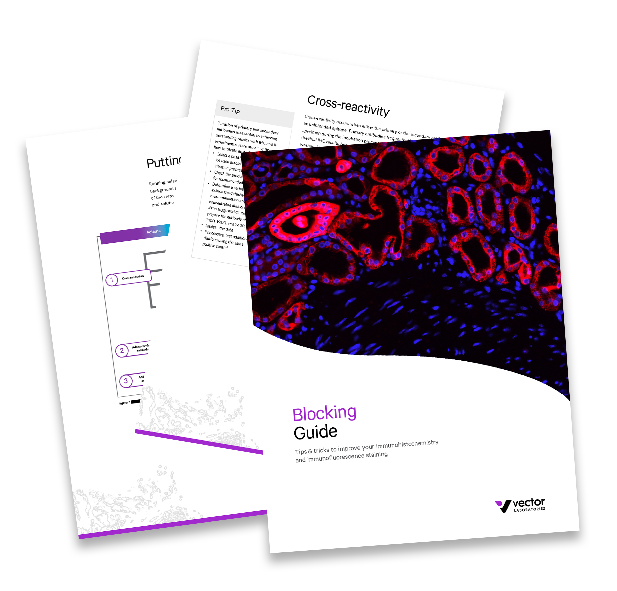
Performing a successful immunohistochemistry (IHC) or immunofluorescence (IF) experiment requires the perfect combination of primary antibody, secondary antibody, and detection system. Blocking is a key step to ensure low background and a clear staining of the target epitope. When performed correctly, the blocking step reduces non-specific binding and quenches autofluorescence and endogenous enzyme activity, resulting in clear visualization of the target epitopes. In this guide, we will walk you through practical solutions to each potential source of non-specific staining in IHC and IF applications to empower you to improve your staining results.